Preface
I first encountered PDRN at a Korean dermatology conference in Seoul three years ago. A clinician presented split-face data showing that one side treated with a PDRN skin booster outperformed the HA filler side at every follow-up — week 4, week 16, and week 28. The room was quiet. When I asked the presenter which salmon species their PDRN was sourced from, she paused and said, "We trust the manufacturer on that."
That moment crystallized something for me: PDRN had the clinical evidence, but its supply chain was a black box that few formulators understood.
This article is not a marketing narrative about "salmon DNA miracles." It is a technical walkthrough of where PDRN actually comes from — the species, the tissue, the extraction chemistry, the quality benchmarks. Because when you are formulating a product or building a brand around regenerative ingredients, you need to know what you are buying, not just what the brochure says.
The Source: Why Salmon and Trout?
Oncorhynchus keta and Oncorhynchus mykiss
!Comparison of chum salmon and rainbow trout as PDRN source species
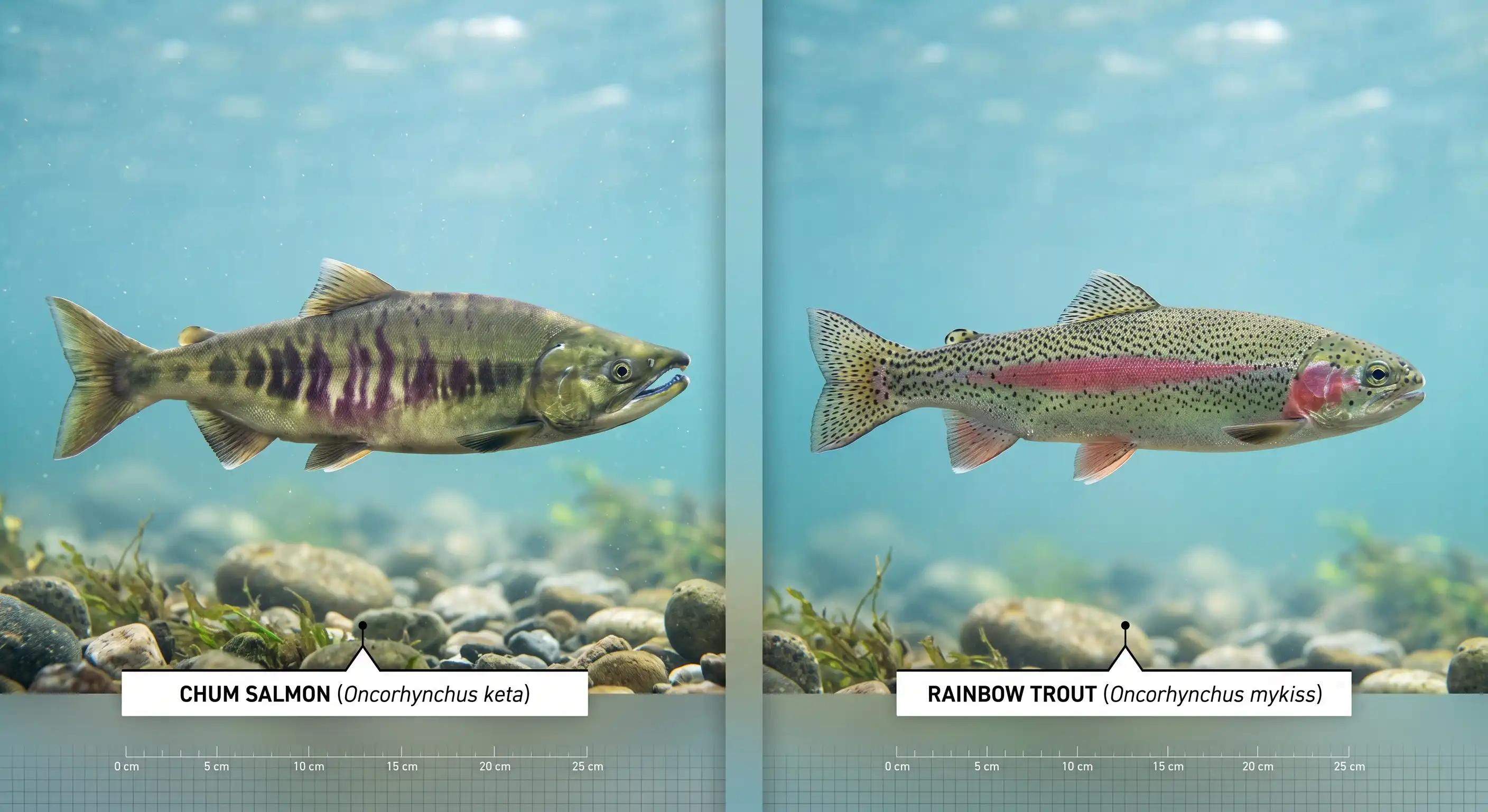
PDRN is extracted primarily from two fish species:
| Species | Common Name | DNA Content (wet tissue) | Primary Use |
|---|---|---|---|
| Oncorhynchus keta | Chum salmon | ~7.5% | Korean PDRN products (Rejuran, Nucleofill) |
| Oncorhynchus mykiss | Rainbow trout | ~7.5% | European pharmaceutical PDRN (Placentex) |
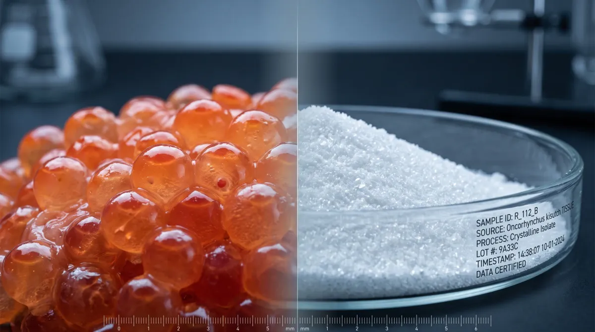
Salmon and trout were not chosen arbitrarily. Their sperm cells — specifically the milt (semen) collected from mature males — have the highest DNA density of any readily available biological tissue. In adult salmon testes, DNA represents approximately 7.5% of the wet tissue weight [1]. To put that in perspective: mammalian liver tissue, a standard DNA extraction source in molecular biology, contains roughly 0.2–0.5% DNA by wet weight. Salmon sperm is 15 to 35 times more DNA-dense.
This exceptional DNA density is a consequence of reproductive biology. Salmon sperm cells are specialized biological delivery vehicles: they contain a tightly compacted haploid genome with minimal cytoplasm and negligible organelle content. A single salmon sperm cell is essentially a DNA payload with a propulsion tail — evolution optimized for genetic delivery, not cellular complexity. This means the starting material for PDRN extraction is already approximately 60–65% DNA by dry weight in the nuclear fraction, compared to less than 5% for typical mammalian somatic cells [1].
Why Not Other DNA Sources?
PDRN could theoretically be extracted from any organism — all living cells contain DNA. The question is why salmon dominates commercially:
1. Extraction efficiency. The high DNA-to-protein ratio means fewer purification steps, lower solvent use, and higher yield per kilogram of starting material. This translates directly to cost per gram of finished PDRN.
2. Low immunogenicity. Vertebrate DNA — including salmon and trout DNA — has inherently low immunogenicity in mammals. This is due to CpG suppression: bacterial DNA contains frequent unmethylated CpG dinucleotide motifs that activate mammalian Toll-like receptor 9 (TLR9) as an innate immune danger signal. Vertebrate DNA has a significantly lower CpG frequency and typically carries methylation marks, making it immunologically "quiet" [2].
3. Established regulatory precedent. Because PDRN was first approved as a pharmaceutical product (Placentex® in Italy, 1994) using trout DNA, the regulatory pathway for salmonid-sourced PDRN is well-defined. Using a novel source organism would require new toxicology and safety dossiers — a multi-year, multi-million-dollar undertaking.
4. Supply chain maturity. The commercial fishing industry for Pacific salmon (particularly chum salmon in Korea and Japan) generates milt as a byproduct. Using this byproduct stream for pharmaceutical and cosmetic extraction adds value without requiring dedicated fish harvesting.
The Extraction Process: From Milt to White Powder
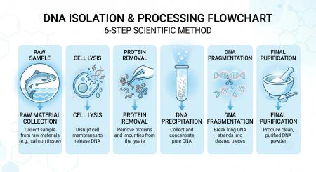
The industrial production of cosmetic-grade PDRN follows a multi-step protocol that transforms raw salmon milt into purified DNA powder. While proprietary variations exist between manufacturers, the core process chemistry is well-documented [1][3]:
Step 1: Raw Material Collection
Salmon milt is collected from mature male fish during the spawning season. Collection is typically performed at commercial fish processing facilities. Milt is immediately frozen or processed to prevent enzymatic degradation of DNA by endogenous nucleases. Freshness of the starting material is critical — DNA in post-mortem tissue degrades rapidly if not stabilized.
Step 2: Cell Lysis
Frozen milt is thawed and homogenized. Sperm cell membranes are disrupted using a combination of mechanical shear and chemical lysis. The goal is to release nuclear DNA into solution while minimizing mechanical shearing that would prematurely fragment the DNA.
Step 3: Protein Removal
This is the most critical purification step. Proteins — including histones (the DNA-packaging proteins), protamines (sperm-specific DNA-binding proteins), and residual enzymes — must be quantitatively removed. This is typically achieved through:
- Enzymatic digestion: Protease treatment (e.g., proteinase K) degrades protein contaminants into soluble peptides
- Chemical precipitation: Salting-out or organic solvent precipitation selectively removes proteins while keeping DNA in solution
- Phase separation: Phenol-chloroform extraction (traditional) or detergent-based phase separation (modern) partitions proteins into the organic phase while DNA remains in the aqueous phase
The goal is protein content below 5% of the final dry weight, with no detectable DNase or RNase activity.
Step 4: DNA Precipitation and Recovery
DNA is precipitated from the aqueous phase by adding ethanol to 70% final concentration in the presence of sodium acetate (0.3 M). The precipitated DNA forms a white, fibrous mass that is collected by centrifugation or spooling. This step also removes residual salts and small-molecule contaminants.
Step 5: Controlled Fragmentation
Intact salmon genomic DNA is enormous — chromosome-sized molecules of hundreds of millions of base pairs. For PDRN, the DNA must be reduced to the therapeutically relevant size range of 50–2,000 base pairs (approximately 50–1,500 kDa molecular weight) [1].
Fragmentation is achieved through controlled enzymatic or physical methods:
- Enzymatic: Controlled DNase I digestion with timed incubation
- Physical: Ultrasonic shearing (sonication) at calibrated power and duration
- Thermal: Heat treatment under controlled conditions (dry DNA is stable below 190°C)
The resulting fragment size distribution is verified by agarose gel electrophoresis or HPLC size-exclusion chromatography.
Step 6: Final Purification and Drying
The fragmented PDRN solution undergoes final filtration through 0.45 μm and 0.20 μm membranes to remove particulates and ensure sterility. The purified solution is then lyophilized (freeze-dried) to produce the final product: a white to off-white powder or fibrous solid that is water-soluble and stable at room temperature.
Purity and Quality: What the Spec Sheet Means
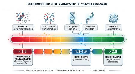
When evaluating a PDRN Certificate of Analysis (COA), the following parameters are critical [1][3]:
DNA Purity: OD 260/280 Ratio
The UV absorbance ratio at 260 nm (DNA) versus 280 nm (protein) is the universal purity indicator. Pure DNA has an OD 260/280 ratio between 1.8 and 2.0.
- >2.0: Possible RNA contamination
- 1.8–2.0: Pure DNA — acceptable for cosmetic and pharmaceutical use
- 1.6–1.8: Moderate protein contamination — requires additional purification
- <1.6: Significant protein contamination — not suitable for PDRN applications
For reference, the L-PDRN study reported an OD 260/280 ratio of 1.91 for their microbial PDRN [4].
Molecular Weight Distribution
PDRN is not a single molecular species — it is a mixture of DNA fragments with a defined size range. The specification should state:
- Target range: 50–2,000 bp (base pairs)
- Corresponding molecular weight: 50–1,500 kDa
- Verification method: Gel electrophoresis with DNA ladder standard or HPLC-SEC
Fragments below 50 bp are too small to effectively engage the A2A receptor and are rapidly degraded. Fragments above 2,000 bp have poor tissue penetration and slower enzymatic processing to release adenosine.
pH and Solubility
- pH: 6.0–8.0 in aqueous solution
- Solubility: Freely soluble in water; insoluble in organic solvents
- Appearance: White to off-white powder; solution should be clear and colorless
Heavy Metals and Microbial Limits
- Heavy metals (Pb, As, Hg, Cd): Below ICH Q3D or equivalent pharmacopoeia limits
- Total aerobic microbial count (TAMC): ≤100 CFU/g (cosmetic grade)
- Total yeast and mold count (TYMC): ≤10 CFU/g
- Absence of specified pathogens: E. coli, Salmonella, Pseudomonas aeruginosa, Staphylococcus aureus
Residual Solvents
If organic solvents are used in extraction, residual levels must comply with ICH Q3C or USP <467> guidelines. Modern aqueous-based extraction methods increasingly eliminate organic solvents entirely, reducing this concern.
Cosmetic Grade vs. Pharmaceutical Grade PDRN
The distinction between cosmetic-grade and pharmaceutical-grade PDRN is not formally defined in most pharmacopoeias, but industry practice recognizes two tiers [1][3]:
| Parameter | Cosmetic Grade | Pharmaceutical Grade |
|---|---|---|
| DNA Purity (OD 260/280) | ≥1.8 | ≥1.9 |
| Protein Content | <5% | <2% |
| Endotoxin | Not specified (typically tested) | <0.5 EU/mg |
| Sterility | Microbial limits per ISO 17516 | Sterile (per Ph. Eur. / USP) |
| Fragment Size Distribution | 50–2,000 bp | 80–1,500 bp (tighter range) |
| Residual Solvents | Per ICH Q3C | Per ICH Q3C |
| Batch-to-Batch Consistency | Acceptable variation | Tight specification range |
| Regulatory Pathway | Cosmetic ingredient (INCI: sodium DNA) | Medical device or drug product |
| Cost | $–$$ | $$$–$$$$ |
For topical cosmetic formulations, cosmetic-grade PDRN is appropriate and cost-effective. For injectable skin boosters, mesotherapy products, or products making therapeutic claims, pharmaceutical-grade PDRN is required to meet the stricter regulatory standards for parenteral products.
The Vegan Alternative: L-PDRN from Lactobacillus
In January 2025, a research team from Chungbuk National University and MNH Bio published the first characterization of microbial-derived PDRN [4]. Their approach replaces salmon milt with Lactobacillus rhamnosus — a GRAS (Generally Recognized as Safe) probiotic bacterium isolated from wild soybean (Glycine soja) native to Korea.
How L-PDRN Is Made
The extraction process is conceptually similar but adapted for bacterial cells:
- Fermentation: L. rhamnosus is cultured in MRS medium at industrial scale (10^10–10^11 CFU/g)
- Cell harvesting: Centrifugation, washed with saline to remove extracellular components
- Heat lysis: 121°C, 0.2 MPa — simultaneous sterilization and DNA release
- Purification: Filtration (0.45 μm → 0.20 μm), ethanol precipitation, washing, dissolution in TE buffer
The process is entirely chemical-solvent-free, using only thermal and mechanical methods for cell disruption.
How L-PDRN Compares to Salmon PDRN
| Characteristic | Salmon PDRN | L-PDRN (Microbial) |
|---|---|---|
| DNA Fragment Size | 200–800 bp | <100 bp |
| OD 260/280 | 1.8–2.0 | 1.91 |
| Antioxidant Activity (DPPH, 5000 ppm) | 8.45% | 40.78% (4.8× higher) |
| Cell Protection (H₂O₂ stress, 500 μg/mL) | 72.43% viability | 82.31% viability |
| Wound Healing (inflammatory conditions) | Baseline | Significantly faster |
| A2A Receptor Pathway | ✅ Yes | ✅ Yes (confirmed) |
| Additional Pathways (p38, ERK) | ❌ No | ✅ Yes (unique) |
| Human Clinical Data | 20+ years, multiple RCTs | None |
| Production | Seasonal, resource-limited | Year-round, scalable |
| Sustainability | Byproduct valorization | Fermentation (low footprint) |
| Vegan | ❌ No | ✅ Yes |
L-PDRN's most notable advantage is its smaller molecular weight: DNA fragments below 100 bp are expected to penetrate the stratum corneum more effectively than the 200–800 bp fragments in salmon PDRN — potentially improving topical delivery without formulation tricks [4].
However, the elephant in the room is clinical evidence. Salmon PDRN has 20+ years of human data, including double-blind RCTs with hundreds of patients. L-PDRN has in vitro data on HaCaT keratinocytes and Raw264.7 macrophages — valuable, but not equivalent. For brands making evidence-based claims, salmon PDRN remains the gold standard. For brands targeting the vegan and sustainability-conscious market, L-PDRN offers a credible science-backed alternative that will likely accumulate clinical data in the coming years.
Sourcing PDRN: What Formulators Should Ask Suppliers
When evaluating a PDRN supplier, request and review the following documentation:
- Certificate of Analysis (COA): DNA purity (OD 260/280), protein content, pH, appearance
- Molecular Weight Distribution: Gel electrophoresis profile showing fragment size range
- Heavy Metal Analysis: ICP-MS or equivalent, per ICH Q3D
- Microbial Limits: TAMC, TYMC, pathogen screening
- Species Origin: O. keta or O. mykiss — impacts regulatory pathway
- Manufacturing Flow Chart: Process overview including sterilization steps
- Stability Data: Shelf life under recommended storage conditions
- Regulatory Support: DMF (Drug Master File) availability, INCI registration status, country-specific compliance documentation
GINKVORA supplies cosmetic-grade salmon-derived PDRN powder with full documentation support. Contact our team for COA, TDS, and sample availability.
Related Articles
- What Is PDRN? The Complete Guide to Salmon DNA in Skincare — The companion article covering PDRN mechanism, clinical evidence, and skincare applications
- Glabridin: Licorice Extract vs Purified Glabridin 98% — Another ingredient where source material purity directly affects cosmetic performance
- EGCG 98% Purity vs Green Tea Extract 50% Polyphenols — How ingredient specification and purity tiers affect formulation outcomes
References
Lee JH, Han JS, Kong HH, et al. Recent Advances on Polydeoxyribonucleotide Extraction and Its Novel Applications in Cosmeceuticals. International Journal of Biological Macromolecules. 2024;283:137852. https://doi.org/10.1016/j.ijbiomac.2024.137852
Veronesi F, Dallari D, Sabbioni G, et al. Polydeoxyribonucleotides (PDRNs): From Physical Chemistry to Biological Activities and Clinical Applications. International Journal of Molecular Sciences. 2017;18(9):1927. https://doi.org/10.3390/ijms18091927
Squadrito F, Bitto A, Irrera N, et al. Pharmacological Activity and Clinical Use of PDRN. Frontiers in Pharmacology. 2017;8:224. https://doi.org/10.3389/fphar.2017.00224
Seo WS, Kang DJ, Chae DB, et al. First Report on Microbial-Derived Polydeoxyribonucleotide: A Sustainable and Multifunctional Alternative to Salmon PDRN. Current Issues in Molecular Biology. 2025;47(1):41. https://doi.org/10.3390/cimb47010041