
TL;DR
EGCG inhibits all three collagenases that UV radiation activates in human skin — MMP-1, MMP-8, and MMP-13 — at concentrations achievable in topical cosmetic formulations (0.5–2.0%). This is not generic antioxidant activity. It is direct enzyme inhibition combined with upstream signaling blockade at AP-1 and NF-κB. Most formulators think of EGCG as "green tea antioxidant." That description misses the mechanism entirely. Here is the before-and-after data, the molecular pathway, and what the numbers mean for someone building a photoprotection product.
What UV Does to Collagen in 72 Hours
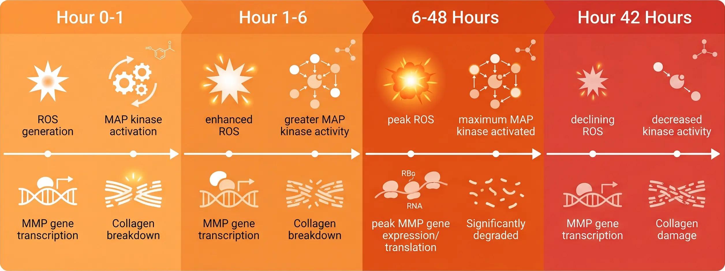
The timeline matters. Thirty minutes of midday sun exposure does not visibly age skin on the spot. What it does is initiate a biochemical chain reaction that runs for days:
Hour 0–1: UVB photons (290–320 nm) penetrate the epidermis and reach the papillary dermis. Energy absorption by chromophores — primarily trans-urocanic acid and tryptophan residues — generates reactive oxygen species (ROS). Within minutes, ROS levels in irradiated fibroblasts spike.
Hour 1–6: ROS act as second messengers, activating the MAP kinase cascade. Phosphorylated JNK and p38 MAPK accumulate in the cytoplasm, then translocate to the nucleus. c-Jun and c-Fos — the two components of transcription factor AP-1 — are phosphorylated and dimerized. Simultaneously, IκBα undergoes phosphorylation and proteasomal degradation, releasing NF-κB for nuclear translocation.
Hour 6–48: AP-1 and NF-κB bind to promoter regions of MMP genes. MMP-1 (collagenase-1) mRNA transcription increases 5- to 10-fold in human dermal fibroblasts exposed to 30 mJ/cm² UVB — a dose equivalent to roughly 20 minutes of midday summer sun at temperate latitudes. MMP-8 (collagenase-2, neutrophil collagenase) and MMP-13 (collagenase-3) follow a similar induction profile. Among these, MMP-1 is the most clinically significant because it is the only mammalian enzyme capable of cleaving intact, triple-helical type I collagen at the single Gly-Ile/Leu bond in the α1 chain.
Hour 48–72: MMP-1 protein reaches peak concentration in the dermal extracellular matrix. The enzyme binds to type I collagen fibrils and makes the initial cleavage. Once the triple helix is unwound at the cleavage site, MMP-2 and MMP-9 (gelatinases) process the denatured collagen fragments. The result is a net loss of dermal collagen density — a microscopic event that, repeated hundreds of times over years, becomes visible as wrinkles.
This is the cascade. Any intervention point in this chain has the potential to reduce the final collagen loss. EGCG intervenes at three points simultaneously.
Where EGCG Intervenes: Three Targets, One Molecule
The gallate moiety is what makes EGCG different from every other green tea catechin. EC (epicatechin) lacks it. EGC (epigallocatechin) lacks it. ECG (epicatechin gallate) has it but lacks the B-ring hydroxylation pattern that gives EGCG its superior radical-scavenging capacity. Only EGCG combines the trihydroxy B-ring with the gallate ester at C-3 — and both structural features are independently required for its anti-collagenase activity.
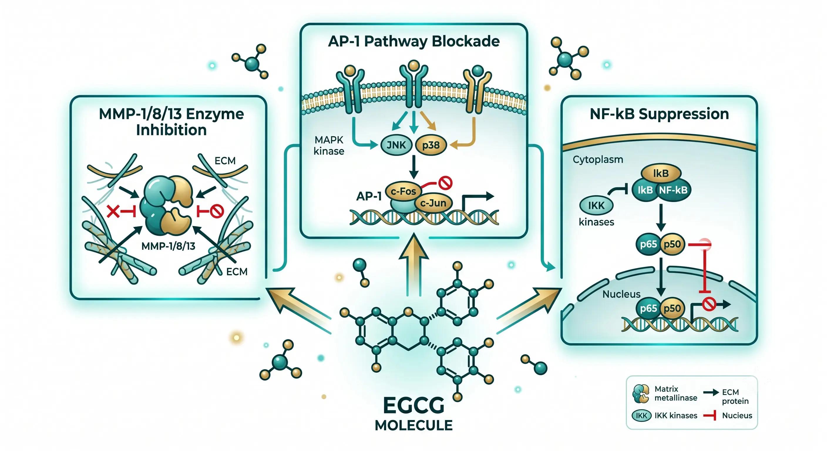
Target 1: Direct MMP Inhibition
The catalytic domain of MMP-1 contains a zinc ion coordinated by three histidine residues in the conserved HExxHxxGxxH motif. The gallate moiety of EGCG chelates this catalytic zinc, physically blocking substrate access to the active site. This is not a signaling effect — it is stoichiometric enzyme inhibition. Pre-treatment of human dermal fibroblasts with EGCG at 12.5 μg/ml before UVB exposure reduced MMP-1 protein secretion and mRNA transcription levels, with the effect mediated partly through JNK pathway inhibition Bae et al., 2008, Food and Chemical Toxicology.
The same study demonstrated that EGCG dose-dependently inhibited not just MMP-1 but also MMP-8 and MMP-13 production, confirming that the effect spans all three major dermal collagenases. No other single natural molecule has been shown to inhibit all three UV-induced collagenases at topical-formulation-achievable concentrations.
Target 2: AP-1 Pathway Blockade
UVB exposure triggers phosphorylation of c-Jun via JNK (c-Jun N-terminal kinase) and phosphorylation of c-Fos via ERK (extracellular signal-regulated kinase). The phosphorylated heterodimer forms the active AP-1 transcription factor complex, which binds to the TRE (TPA-responsive element) in MMP gene promoters.
EGCG suppresses both JNK and ERK phosphorylation, thereby reducing the pool of active c-Jun and c-Fos available for AP-1 complex formation. A 2024 study by Zhang, Xu, and colleagues demonstrated this mechanism in both zebrafish and human skin fibroblast (HSF) models: EGCG pretreatment reduced UVR-induced phosphorylation of p38 MAPK and suppressed downstream NF-κB and AP-1 activation Zhang & Xu et al., 2024, Mediators of Inflammation.
The key insight: EGCG does not merely scavenge the ROS that trigger AP-1 — it directly interferes with the kinase enzymes that execute AP-1 activation.
Target 3: NF-κB Suppression
NF-κB is the master regulator of inflammation. UVB triggers IκB kinase (IKK) activation, which phosphorylates IκBα, tagging it for ubiquitination and proteasomal degradation. Once freed from IκBα inhibition, NF-κB (p50/p65) translocates to the nucleus and activates transcription of pro-inflammatory cytokines (IL-1β, TNF-α, IL-6) as well as MMP genes.
EGCG inhibits IKK activity, preserving IκBα and keeping NF-κB sequestered in the cytoplasm. This breaks the positive feedback loop between inflammation and collagen degradation: less NF-κB → fewer inflammatory cytokines → less secondary MMP induction → less collagen loss. The same 2024 Zhang & Xu study confirmed that NF-κB pathway suppression is a core component of EGCG's photoprotective mechanism, alongside p38 MAPK and AP-1 regulation.
Why These Three Targets Matter Together
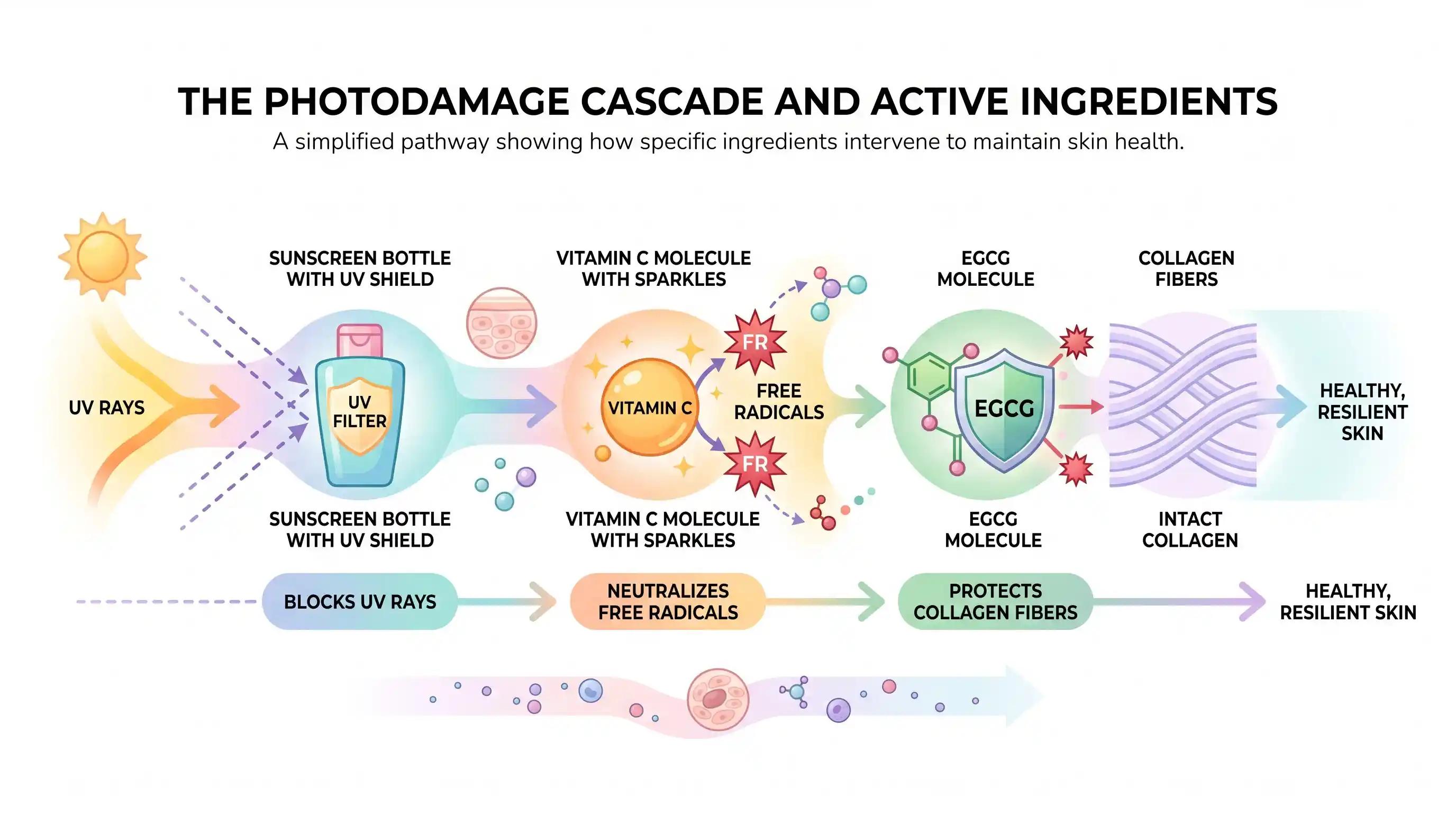
The three mechanisms operate on different time scales: Direct MMP inhibition is immediate (minutes, enzymatic), AP-1 and NF-κB blockade are transcriptional (hours, gene expression), and the combined effect reduces collagen degradation at every stage of the UV response cascade. A single ingredient that only scavenges ROS — like vitamin C — operates only at the initiation stage. A single ingredient that only inhibits one MMP — like a synthetic MMP inhibitor — misses the upstream signaling that drives ongoing collagenase production. EGCG is one of very few natural molecules that addresses the full chain.
The Before-and-After Evidence
 The mechanistic data is strong, but here is the evidence that matters for a product claim:
Human skin in vivo. A controlled study of 18 volunteers (age 21–71) applied topical green tea extract before UV exposure and measured sunburn cell formation against a placebo-treated control site. Green tea extract pretreatment produced a significant reduction in sunburn cells. A concentration gradient from 0.25% to 10% was tested: 0.5% showed measurable photoprotective activity, and 2.5% provided what the investigators described as "excellent protection" cited in MDPI Molecules, 2024, 29(22):5226.
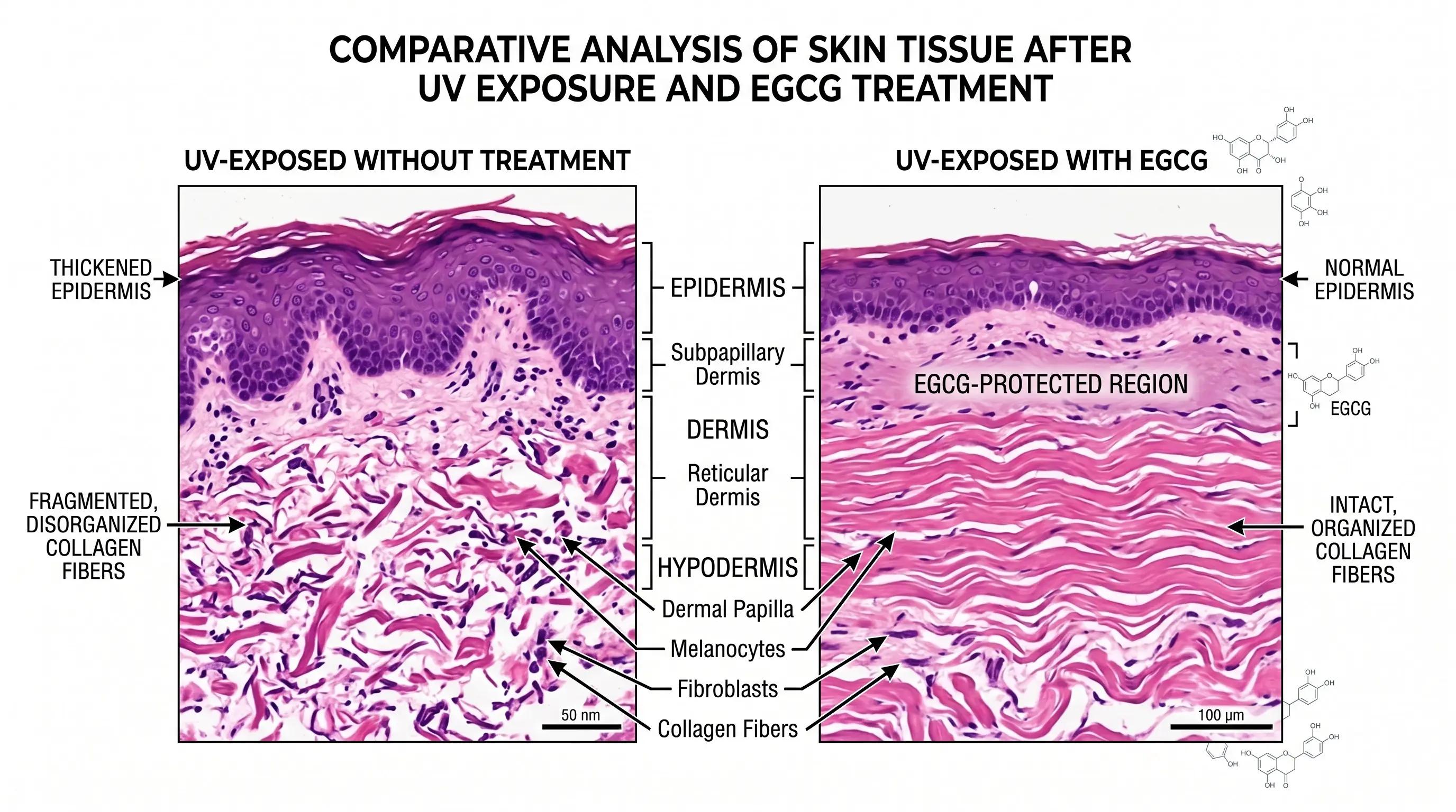
Human skin ex vivo. A 2024 structure-based virtual screening study found that in a human skin tissue model, EGCG prevented UV-induced collagen breakdown and preserved normal epidermal thickness. The effect was visible histologically — irradiated skin without EGCG showed fragmented collagen fibers and epidermal hyperplasia, while EGCG-treated irradiated skin maintained collagen fiber integrity and normal epidermal architecture Heliyon, 2024, 10(21):e40373.
Human dermal fibroblasts in vitro. EGCG at 12.5 μg/ml reduced MMP-1 protein secretion and mRNA expression in HSF cells exposed to 30 mJ/cm² UVB. The inhibitory effect was consistent across both UVA- and UVB-induced MMP-1 upregulation, indicating that EGCG's photoprotective mechanism is wavelength-independent — it works against the biological response regardless of which UV band initiates it Bae et al., 2008.
A 2023 study confirmed that EGCG-treated UVA-exposed human skin fibroblasts showed reduced MMP-1 and MMP-3 expression with a corresponding increase in type I collagen and procollagen expression Jia et al., cited in MDPI Antioxidants, 2024, 13(12):1506.
Zebrafish model. In the 2024 Zhang & Xu study, EGCG pretreatment of UVR-exposed zebrafish significantly reduced skin wrinkling and collagen degradation compared to untreated controls. The zebrafish model is particularly informative because zebrafish skin shares structural and molecular features with human skin, including a stratified epidermis and a collagen-rich dermal layer, but the organism is transparent — allowing direct visualization of collagen structure without histological sectioning.
Animal model. Mice fed green tea polyphenols before chronic UV exposure showed significantly higher dermal hydroxyproline content (a quantitative marker of collagen density), increased catalase activity (indicating enhanced endogenous antioxidant defense), and decreased protein carbonyl content (a marker of oxidative protein damage). Hydroxyproline is a specific and quantitative collagen marker because it is almost exclusively found in collagen and represents approximately 13.5% of collagen by weight cited in MDPI Molecules, 2024.
Meta-review level. A 2019 review published in Nutrients examined 21 studies (6 in vitro, 9 review papers, 6 controlled human trials) on green tea catechins and skin health. The conclusion: consistent evidence for anti-wrinkle activity, increased cell proliferation, and reduced oxidative damage from both topical and oral green tea catechin administration. No serious adverse events were reported in any of the human studies at the concentrations used.
What This Means for Your Formula
If you are building a photoprotection or anti-aging product, here is what the data translates to in practical formulation terms:
The concentration math. Human data shows photoprotection starting at 0.5% EGCG. If you are using a standard "50% polyphenols" green tea extract — which contains roughly 15% EGCG by weight — you need 3.3% extract in your formula to reach the minimum effective EGCG concentration. At 3.3%, you are also adding ~0.1% caffeine, ~1.7% other catechins with unknown interactions, and a brown-green color that requires masking. If you are using EGCG 98% (HPLC-purified), you need 0.51% to reach the same effective EGCG dose — with no caffeine, no competing catechins, and significantly less color impact.
The stability reality. EGCG degrades rapidly at pH above 5.0. If your photoprotection serum is formulated at pH 5.5–6.5 for skin compatibility, unprotected EGCG will oxidize before the product reaches the consumer. I have written a separate, detailed stability protocol that covers the five variables you need to control — pH buffering, antioxidant pairing (ascorbic acid at 0.05–0.1%), oxygen exclusion (nitrogen flushing + vacuum emulsification), light-protective packaging, and chelating agents (EDTA or phytic acid at 0.05–0.1%). Read the full stability guide here.
The photoprotection ≠ sunscreen distinction. EGCG does not have an SPF value. It does not absorb UVB photons in the 290–320 nm range at levels that would meaningfully reduce UV dose at the skin surface. What it does is neutralize the biological consequences of the UV that penetrates your skin regardless of sunscreen application. Even SPF 50 allows 2% of UVB to reach the epidermis. Over decades, that 2% drives cumulative collagen loss. EGCG functions as a biological backstop — it reduces the collagenase response to the UV that gets through.
The synergy opportunity. If your formula already contains a UV filter and vitamin C, adding EGCG adds a dimension neither ingredient provides: enzyme-level collagen protection. Vitamin C scavenges ROS (pre-MMP induction), UV filters block photons (pre-ROS generation), and EGCG inhibits MMP catalytic activity and transcriptional signaling (post-ROS, pre-collagen cleavage). These three ingredients target three sequential stages of the photodamage cascade.
The evidence threshold for claims. The data supports claims around "collagen protection," "UV-induced enzyme inhibition," and "photodamage defense." It does not support SPF-equivalent claims or DNA repair claims — EGCG's mechanism is collagenase-focused and inflammation-focused, not genome-maintenance-focused. The strongest evidence base is at the MMP-1/AP-1/NF-κB intersection, and claims should reflect that specificity.
Related Articles
- EGCG Formulation Stability Guide — The five variables that control EGCG degradation in cosmetic and nutraceutical formulations
- Liposomal Delivery Technology Hub — Why encapsulation matters for unstable molecules like EGCG
- Quercetin Senolytic Anti-Aging — Another plant polyphenol with multi-target anti-aging mechanisms